My British Osteological Database – An Update

skele_1

Finding research material is major component for any PhD. This can often mean extensive online searchers, emails to curators and searching through journal articles for relevant or useful material.

From my own experiences (where I needed large samples of human molars from a range of archaeological periods) this is not always an easy thing to do. A while back I decided to produce a crude database about some of the human remains collections I came across during my search. Recently, I decided to update this to make it more user friendly and to add a couple more sites.

You can find a downloadable excel file here with site names, dates and basic collection information.

Enjoy!

PS. If there are any more collections that you know of (and their information can be shared publicly) please get in touch!

Oops I Did it Again…

oops i did it again memeI’ve left it a while since writing a blog post! Sorry the PhD took over my life for a while there (a bit more than usual!).

So what have I been doing? Well there has been some more data collecting, more writing, more hockey and the upgrade. For those of you who don’t know about the PhD process some universities conduct upgrades during the PhD. This is an examination type meeting where a couple of lecturers (most likely within the same department as you) question the PhD student on a piece of written work and assess whether they a) have a project that can stand and process onto a PhD and 2) that the student knows what they are doing?!

To be honest mine feels like a bit of a blur and it is quite an odd experience! However, I came through it and received some great advice that I will be using to refine, restructure and focus my project. I’m hoping that I’ll continue my PhD with a slightly new perspective and with the knowledge that it is going well (although I will have to remind my self that constantly. It’s the nature of the beast!).

Please note that for each university this process is different and has different requirements. When I talk about my upgrade it only relates to my experience. I highly recommend attending any training events relating to the upgrade and/or talk to your supervisors and fellow students who have gone through it. I was told that this is to help prepare you for the final viva so it is going to be tough – but worth it in the end! 

So what else have I been doing? Well more data collection for a start. I’ve visited a couple more museums including Cheltenham Museum and a small collection held by the University of Bristol Spelaeologial Society (which also hosts the brilliant cave Gazetteers website that I mention on my British Osteologial Collections page). But I guess the one I would like to brag about is the Natural History Museum! Yup I was lucky enough to carry out a weeks data collection at the NHM London.

The NHM is one of my favourite museums, I went there a number of times and now that I live near London I can go as often as I like. But it was such a great opportunity to go and use some of their collection in my research. The lovely Curatorial Assistant was on hand to help with any questions and queries and it was great getting to chat to her. I hope to see her again at BABAO 2017 where we both hope to present a poster. It was a privilege to work there and can even say that I met the curator of the human remains collections (who was also our first key note speaker at our SSSB conference – see previous post). In addition, as part of the agreement to collect data I carried out some basic osteological curation work on the specimens I examined by completing an inventory sheet of the remains. It may sound like a small thing but it will allow for easier and more efficient inventorying and assessment for future researches and curators – and hey every little helps!

What else is there…Oh yeah I’ve just come off from two excellent training days organised by the Collaborative Doctoral Partnership (CDP), who fund my PhD. These two days talked about life after the PhD, getting and applying for funding grants and things to consider when applying for jobs. Even though I am only in my 2nd year to was a great thing to attend. It means I can start thinking about what I want to do now, and start preparing my CV, skills and research so I can look for work/funding efficiently and productively when the time comes. It was also great to meet some of the other CDP students, it’s always nice to hear about their experiences and learn about their research. Through this I was asked if I would like to do a small presentation about object handling at another CDP event coming up in a few weeks (check out those networking skills lol!). They haven’t got anyone talking about human remains so I think I will take them up on their offer. Plus it will give me another chance to practice my presentation skills! I’ll report back in a few weeks to let you know how it went!

Anyway, I hope this gives you a little idea of what I’ve been up to. Next week I’m away in Barcelona (it’s the boyfriend’s birthday treat) and will come back refreshed and relaxed ready to hit the ground running with the PhD. Until then…adios!


Return to Cardiff Museum

Sorry I didn’t post anything late week I was rather busy so it slipped my mind, so this post will talk about my last 2 weeks of work. This includes another trip to the stores of the Corinium Museum, volunteering at the Royal College of Surgeons, a trip to the Cotswold Archaeology office and another up to the National Museum of Wales in Cardiff.
My visit to the Corinium Museum stores was successful again. I got through another load of Anglo-Saxon skeletons for my data collection. It’s such a good collection and its going to be very useful for my PhD project as it includes lots of juvenile individuals. This makes me very happy! However, I’ve got quite a few more trips to make to their stores as it’s such a large collection.
The day after my trip to Cirencester I was back at the Royal College  of Surgeons volunteering in the museums department. It’s been a few weeks since  I’ve been there as the museum has been quite busy and haven’t been able to have me in. However, it was great to be back packing more skeletons into boxes ready for the move. Of course, it was also great to see the staff members again. I do enjoy working there!
So that was last week. I started this week by coming up to Kemble, which is near Cirencester. The purpose of this trip was to visit one of the office of Cotswold Archaeology, as they had a couple of Bronze Age and Iron Age skeletons. Whilst there I got to meet a couple of lovely people, including Sharon Clough who gave me some great information regarding some of the other collections I’m intending to use in my research. I also got to chat to Sharon about commercial archaeology units and learn a bit more about them. I’ve only really visited museums and universities so far, so it was a really good opportunity to learn about the commercial sector – a completely new area for me!
This week has ended with a trip but to the National Museum of Wales, in Cardiff. In my last visit I went through the prehistoric human remains in their collection. This time I was going back to the relevant specimens and taking measurements. I’ve now managed to get collection of Neolithic individuals recorded, plus a few Bronze Age remains, which is always good!
So another day, another lot of data collection completed. I think it’s all going well – I feel like I’ve got a lot done, but then I still have a load more to do! As a little fish called Dory once said ‘just keep swimming!’

A Night In An Old Operating Theatre!

This week has been a long one! I’m not sure why as it’s been pretty good and quite productive but it’s taken a while to get through. Maybe it’s because I’ve been travelling for my data collection again and I’m not used to driving  so much?! As well as my PhD work this week I went to a really cool talk about Bodysnatching in an old operating theatre – perfect for Halloween!

On Monday I was back at the stores of the Hampshire Cultural Trust to finish going through the various sites they have. I’m pretty pleased with  myself as I’ve managed to get through a lot of skeletons in a decent amount of time. There are two small sites to work through but as they’ll only take me half a day at most I will return another time. At some point in the future I will need to go to their other store to access a Romano-British population.

On Wednesday I was then back at the stores of Corinium Museum in Cirencester. Although it’s a bit of a journey to get there my mum lives about half an hour away so I went up the night before. This cuts my travel time down in the morning and I get the bonus of seeing my mum. I’m really lucky as I have a great relationship with Mum so it’s always lovely to go back home. Whilst at the Corinium stores I managed to get a decent amount of work done. However, the Anglo-Saxon collection  I’m looking at is quite big. This is good news as it’ll be a great source of data for my PhD but on the other hand it will take me some time to complete it. Unfortunately I can’t do a series of consecutive days at the stores as it is only staffed one day a week, but I then get to visit my mum quite a bit so it’s not all bad!
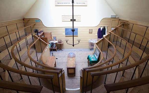
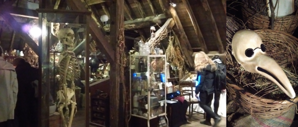
This weeks blog post ends with a trip to the Old Operating Theatre in London to see a lecture. The talk is called ‘Night of the Bodysnatcher’. The Old Operating Theatre is a museum located in the roof space of St. Thomas’s, Southwark just around the corner from London Bridge train station. This is the original site of St Thomas’s hospital and is one of the oldest surviving operating theatres. It is quite an odd place, to access it you have to climb a tight, spiral staircase that leads to a tiny museum displaying some of the instruments and medical equipment used in the past. Going through the museum and around a corner you find the old operating theatre, pictured in the image above. This is where we sat and listened to the talk, but it was strange to think that’s where dissections and operations took place many years ago.
The talk itself was very interesting, I do love learning about the history of surgery and the things surrounding it! This talk was, as the title suggests, about Bodysnatchers or otherwise known as Resurrection Men. These were individuals who took the bodies of the recent dead from their graves and sold them to surgeons, who then used them for dissections to learn about anatomy. It may have been quite a gross job to do, but it could be rather lucrative for a period in the 1700s as surgeons wanted bodies and would therefore pay!
oot

A view of the museum at the Old Operating Theatre, and a replica beak mask.

There were lots of great facts and fascinating bits of information, for example, did you know that the body snatchers stripped the body of corpses of their clothes and possessions and placed them back in the grave? Why – because you could be hung for theft by taking the clothes, as they belonged to the relatives of the deceased, but not for taking the body! It was an incredible insight into the very seedy past of the study of anatomy, and although it was gruesome it allowed many to study the human body. Perhaps without the Bodysnatchers surgery wouldn’t be where it is today! For example, some of the famous early surgeons in the UK, including William and John Hunter, almost certainly would have used snatched bodies in their work!
I very much enjoyed both the talk and museum and I would highly recommend visiting!

The New Plan – Weekly Updates!

So I’ve been a bit rubbish with my blog (again!) but Ive told myself that I am going to be better from now on. I guess I haven’t written much as I haven’t felt that there’s much to say. HOWEVER, I am now back volunteering at the Royal College of Surgeons and I’m going to start visiting museums to access their collections for my PhD work. That means there should be plenty to talk about so I have no excuse for not keeping up with the blog!
So first things first, I’m back at the Royal College of Surgeons volunteering. It’s been about a year since I was last here but I couldn’t help but come back! I enjoyed working here so much and the people are lovely so I was just waiting for the right time.
Now I’m back I’m doing something slightly different then previously, where I was tasked with creating inventories for a couple of collections. This time I’m packing objects/human remains ready for decanting and moving them to a new site. This is because the building in which the museums department is based is getting renovated and so most of the collections have to be moved off site. So far the boxes of remains I have been working on have been straight forward. This has included sorting a couple of boxes of loose ribs and one half (a complete right side) of a skeleton. I’m having to think about how best to wrap and pack each box so that the bones are safe and secure, ready to be moved. As I said the boxes so far have been relatively simple to sort out, but I’m sure there are going to be some tricky ones coming my way!
So that’s my volunteering stuff, now for a quick update about my PhD. I’m now at the stage where I am contacting museums in search of human remains that would be suitable for my project. At times this has been fairly straightforward. I’ve identified a collection, either from some literature or an online resource, then contacted the museum and have been able to find exactly what I am looking for. On other occasions it’s been a little more complicated. For example, I may have found the original excavation report that identifies a collection but it is then difficult to locate the remains. In these situations I have contacted the most likely institutions or commercial archaeological company and went from there. It’s taken a little bit of time but I now feel that I have identified a good amount of skeletal collections, at least as a starting point.
So what’s next? Well I’m visiting the National Museum of Wales in Cardiff and Cirencester museum over the next 2 weeks. Here I  hoping to have a preliminary look at the remains that they have to see how suitable they are for my project. I will (hopefully!) arrange another date to go back and measure any of the remains that can be used in my research. In the mean time I’m going to start contacting the museums that have collections that are appropriate for my work and get some dates for visits in the diary. My aim is to see and measure as many skeletons as possible before April next year, when my PhD upgrade will be. So lots of work to do but I can’t wait to get started with the data collection!
As I said before, now that I’ll be visiting new places and collections, plus the volunteering, I should be able to write an update of my progress and experiences every week! Here’s hoping! Skeleton-Hands-Facebook-Cover

New News!

In the last week two cool things have been confirmed:

I’m really excited about both of these and can’t wait to get stuck in.

The Volunteering

hunterian

Inside the Hunterian Museum. Image taken from here.

About 2 years ago I started volunteering at in the museums department of the Royal College of Surgeons and enjoyed it so much that continued to work there until I started my PhD. Whilst there I was lucky enough to work with some amazing osteology collections and saw some interesting pathologies. I really enjoyed working there and was sad to leave, however, I knew I needed time to settle into my PhD.

A year on I have made the decision to return once a fortnight, so not to impact with my studies too much, to volunteer once again. I am so happy to be returning and to see some of the people I had met previously and can’t wait to get started. I am hoping to start this week, although I am waiting for confirmation, but already know what I will be working on – but I’ll wait until I’ve started to tell you all about it! It will be so lovely to go back, and a positive (and useful!) distraction from my PhD work.

Skeletons, Stories and Social Bodies

SSSB logo

The SSSB logo. Check the conference out here.

A fellow PhD student and friend of mine from Southampton (archaeosarah) and colleagues have set up a new conference called Skeletons, Stories and Social Bodies (SSSB) and I have offered to be a committee member. It will be an inter-disciplinary conference for discussing topics surrounding death, anatomy, attitudes to the body, mortuary practices, and more! This will be a joint conference by the Osteoarchaeology group (Department of Archaeology) and the Centre for Learning Anatomical Sciences (CLAS) at the University of Southampton.

Since volunteering at the Royal College of Surgeons I have become more and more interested in anatomy and therefore saw this as a great opportunity to learn more about the subject. Last year I helped out with the University of Southampton’s student conference, PGRAS, for the archaeology department. I therefore thought helping out with SSSB would be a great way to build on this experience.

Part of my role as a committee member will be to help with the general organisation of the conference and  to read submitted abstracts and proposals. In addition to this I have been asked to help out with promoting the SSSB on social media. This will certainly be a useful skill to develop as so much is carried out in this way now – plus it should help with my networking skills. I’m sure there’ll be lots of other things to help with and I’m definitely going to get stuck in – I may even run a workshop!

Please go and check out the conference and sign up to our mailing list for updates!

Website: http://www.sssbconference.co.uk/

Email: sssbconf@gmail.com

Twitter: @sssbconf or #sssbconf

Facebook: https://www.facebook.com/SSSB2017/


Into the New Year…A PhD Update

My last blog was at Christmas and I’ve been meaning to do another one for the last few weeks but never seemed to get around to it – so here it is finally!

Since the Christmas break (which was lovely and lazy!) and settling back into work I’ve really started to get going with my project. The first step of my project was to propose an adjusted method for recording dental wear in human molars, which was completed before Christmas. This also included identifying the aging method, using human dentition, that would most be the most accurate and easy to apply and came to the conclusion of using the AlQahtani et al (2010) London Dental Atlas. About 2 years ago I wrote another blog piece discussing the accuracy of three different dental age estimate charts, including the London Atlas. The article describes the AlQahtani et al (2010) method in brief but the atlas itself can be viewed here.

The next step was to test out the method that I had created to assess how it worked when practically applied to human skulls. In order to do this I needed a good skeletal collection of the correct time period for my project (neolithic to medieval) that included juvenile and adults skulls. Luckily for me there is such a collection at Southampton and so it was very easy for me to get started! You can find out a bit more about the teaching collection and courses that Southampton University at the following blog: BOS, curated by the wonderful Ellie!

So it’s now the beginning of February and I’ve managed to apply my method to the juvenile skulls twice and the adults once. My plan is to do this a few more times for each set of skulls and there are a few reasons for this. The first is that this will enable to tweek my method where necessary so that it is as easy and accurate to use as possible. The second reason is so that I can become comfortable with handling and identifying teeth, but also so that I get used to the process of recording the data. This will be particularly useful when I move on to different skeletal collections, where I will only have a short period of time with them.

Finally, and probably most importantly, recording the data from the same set of skulls, multiple times will allow me to test the repeatability of my method. In order for this method to be robust and accurate it must produce statistically similar results when applied to the same collection, this will ensure that any results obtained using this method can be comparable. When recording the data from the same collection of skulls I am making sure that there is a period of time in between each data collection. This ensures that I cannot remember any particular individual and all of the collected data is ‘new’ again.

So far, the data collection is going well and I am getting through the skulls in the time that I wanted. Another key aspect of my project is to produce a method that is quick and easy to apply, and at the moment that’s going to plan so fingers crossed it stays that way!

I probably won’t do another blog post on the progress of my PhD for a little while as I won’t have much new to say until I’ve finished my data collection. However, I do plan to do a new blog post on molar identification (seeing as I’m getting pretty good at it now!). Hopefully, I’ll sort that out soon and get it posted!

An Osteological Case of Ancient Down Syndrome.

Skeleton in situ. Taken from Rivollat et al. (2014 Fig. 1 p.9).

Skeleton in situ. Taken from Rivollat et al. (2014 Fig. 1 p.9).

Scrolling through the International Journal of Paleopathology I came across an article entitled ‘Ancient Down syndrome: An osteological case from Saint-Jean-des-Vignes, northeastern France, from the 5–6th century AD’ by Rivollat et al (2014). During my studies I only came across Down Syndrome in genetics it is caused by a partially or complete third copy of chromosome 21, which is why it can be called trisomy 21. I didn’t know much about the physical manifestations of Down syndrome and therefore thought this article might be an interesting read.

Down syndrome is not well documented in human history and only two sub-adult cases have been reported in the archaeological literature. However, it is noted that there is a large amount of variation seen in cases of Down syndrome which makes it difficult to interpret. This study looks at a child’s skull, aged 5-7 years old, from a necropolis dated to 5-6th Century in France. The skull used for study was buried in the same manner as the other skeletons, indicating that it did not have receive different treatment at death.

The individual in question had good bone preservation, although most of the thoracic and lumbar vertebrae and the right hand were missing. This skeleton was originally excavated in 1989, were a photograph was taken in situ (see above). However, post cranial skeleton was lost after this which meant that the current study could only use the skull for analysis. This skull was then compared to 78 skulls of children of similar age and background to look for signs of Down syndrome.

By taking morphometric observations and comparisons some variation of the skull was found. To begin with the cephalic index was larger than the population mean of 83 measuring at 106.2. This is a ration between the maximum width of the head and the maximum length. In the skulls result indicated ‘ultrabrachycrany; i.e.. a skull which was short in length and wider than the norm. This large width of the cranium has been described in Down syndrome patients. The shape of the occipital bone also differs from normal, which is dish shaped, and is flattened in the skull. When observing the sutures the some of the metopic suture was still present and many lambdoid wormian bones were seen. Wormian bones are irregular, isolated bones located within sutures of the cranium, in this case the lambdoid suture which separates the occipital bone from the parietals. These features are also indications of Down syndrome. 

Anterior, inferior and lateral view of the skull, and superior view of the mandible. Taken from Rivollat et al. (2014 Fig. 2 p.9).

Anterior, inferior and lateral view of the skull, and superior view of the mandible. Taken from Rivollat et al. (2014 Fig. 2 p.9).

The mandible was also examined and measured and slight variations from the norm were found. These included a short mandibular symphysis and a narrowing of the breadth of the mandibular ramus. The angle of the jaw, the gonial angle, was slightly above normal. These are features which have been noted by Kisling (1966) and others referenced in the article. 

Another area of interest was the thickness of the skull vault and by using CT scans an accurate measurement could be taken. It was found that the thickness varied over the skull and at anatomical points were inside and outside of the normal range. At the bregma (point at which the coronal suture is intersected perpendicularly by the sagittal suture) and at the naison (intersection of the frontal and two nasal bones) the skull is of normal thickness. At the lambda (meeting point of sagittal and lambdoid sutures) the it is thinner but thicker at the euryon (point on parietal bone marking either end of the greatest transverse diameter of the skull).  The authors claim that the thinness at the back of the skull could be due to poor development of the bone and can be associated with Down syndrome. 

Profile outline of the Saint-Jean-des-Vignes skull and superimposition with another skull of the same age group (O, Opisthion; Ba, Basion; I, Inion; L, Lambda; Br, Bregma; N, Nasion; Ns, Nasospinal; Pr, Prosthion; ST, Sella Turcica). Taken from Rivollat et al. (2014 Fig. 5 p.12).

Profile outline of the Saint-Jean-des-Vignes skull and superimposition with another skull of the same age group (O, Opisthion; Ba, Basion; I, Inion; L, Lambda; Br, Bregma; N, Nasion; Ns, Nasospinal; Pr, Prosthion; ST, Sella Turcica). Taken from Rivollat et al. (2014 Fig. 5 p.12).

For those who are unfamiliar with the anatomical names and points of the skull visit the following web pages. These Bones of Mine provides images and diagrams of the bones of the skull whilst Anatomy Navigator provides a good overview of the anatomical landmarks.

Finally the study looked at the indention of the individual and the authors state that there are many features which are suggestive of Down syndrome. This includes a lack of development of the second lower deciduous premolar, which can affect up to  92% of Down syndrome cases. There is an indication of the onset of peridontal disease by the presence of bone remodelling of the alveolar bone, and which may have caused some tooth loss.

From these features a diagnosis of  Down syndrome was assigned, with a differential diagnosis of rickets and hydrocephalus. However these were ruled out due to the lack of porotic hyoerostosis for rickets and the presence of a normal cranial capacity for the child’s skull. Therefore the skull was given the diagnosis Down syndrome.

Whilst reading this article I did have some concerns about the diagnosis. At the beginning of the article the authors state that there have been few cases of Down syndrome found in the archaeological record, which means they have very few points of comparison. The second issue is that the diagnosis was based solely on the  skull, as the post cranial skeleton was missing.  As I know little about the physical manifestations of Down syndrome I did a quick search and it was very clear that these were very varied. There also appear to be few definite indicators of Down syndrome on the skeleton, and particularly lacking on the skull (this information was indicated from the following website).  taking this into account I think that it was a little unwise to assign the skeleton excavated in the study as having Down syndrome, it may have been more accurate to suggest a possible incidence of the condition.

In fairness to the authors they do state that ‘none of the features is pathognomonic’ (p. 13) however I may have been more conservative in the conclusion, particularly with the absence of the post cranial skeleton.

Rivollat, M., Castex D., Hauret L. & Tillier A. (2014). “Ancient Down syndrome: An osteological case from Saint-Jean-des-Vignes, northeastern France, from the 5–6th century AD.” International Journal of Paleopathology 7(0): 8-14.
Shattered Lives and Broken Childhoods: A Case Study of Child Abuse in the Archaeological Record.

In situ image and schematic of Burial 519. (Fig. 3 page 73). Image taken from Wheeler et al. (2013) article.

In situ image and schematic of Burial 519 (Fig. 3 page 73). Image taken from Wheeler et al. (2013) article.

Recently I have wanted to focus more on human pathologies in archaeology when I came across this article ‘Shattered lives and broken childhoods: Evidence of physical child abuse in ancient Egypt.’ I have never come across an example of this before and therefore gave it a read.

Child abuse is clinically classified as the maltreatment of a child by their parent or caretakers and can include physical, sexual and emotional abuse and physical and/or emotional neglect. In modern cases soft tissue damage and injuries are the most common presentation with 10 – 70% of children showing signs of skeletal trauma. In archaeology it is these latter injuries which may be seen however, it can be difficult to interpret them.

There are many reasons why confusion may occur when attempting to establish child abuse in skeletonised individuals. The first is establishing whether the trauma is a result of an accident or not. Traumas, such as fractures, may look the same no matter how they were obtained. However, the pattern of any pathologies identified, along with their process of healing, may be a good indicator. The use of physical discipline has also been recorded in the archaeology, for example during the Roman Period where it was not uncommon for children to be beaten if they made a mistake. There have been few examples of child abuse found during excavations, which may be a result of poor preservation and preparation, taphonomic processes or adult centred research and therefore does not mean that child abuse didn’t occur in past human societies, only that few cases have been confirmed.

This study by Williams et al. (2013) looks at an individual aged between 2 and 3 years old from the Romano-Christian Period from a cemetery in the Dakhleh Oasis, Egypt. In total 770 individuals were excavated, with a possible 4000 burials being present as indicated by an archaeological survey. From these 158 were 0 -1 years old, including the individual chosen for study. Burial 519 was undisturbed and had all of their teeth and bones, the preservation was also very good and therefore some hair, skin and nails survived. This individual was buried in the same manner as the other juveniles within the cemetery and it’s location did not distinguish it as being atypical. In order to study the individual full radiographs, micro-CT scans and tissue samples were taken, in addition to the standard ontological observations.

Burial 519 was aged using standard ageing methods by identifying the dental development and epiphyseal fusion of the individual. It was also observed that cribra orbitalia (an indication of a pathological deficiency) could be seen in both orbits. The authors noted that 60% of 1-3 year olds at the cemetery had this condition, with 95% of individuals having active lesions. The remaining pathologies recorded for the individual in Burial 519 related to fractures, a full summary of which can be found in Table 1, page 75 of the article.

image showing fractures and bone growth in the humerus and ribs (Fig. 5 p. 76). Image taken from Wheeler et al. (2013) article.

Image showing fractures and bone growth in the humerus and ribs (Fig. 5 p. 76). Image taken from Wheeler et al. (2013) article.

These fractures were found in both upper arm bones, the clavicle, two ribs, two vertebrae and the pelvis. The healing process of these injuries differ and therefore suggests that they occurred at different times. For example, both humeri have complete transverse fractures of the proximal third where the bone margins are slightly rounded and the trabecular bone has a smooth appearance. This indicates that the breaks occurred several weeks before death whilst the fractures of the 7th left and 8th right ribs are well healed. In contrast to these breaks the right clavicle has a complete transverse fracture where there are no signs of healing. From looking at these sites alone it is fairly clear that all of the injuries did not occur at the same time.

This pattern of fractures and healing is consistent with clinical pattern of skeletal trauma in victims of non-accidental trauma; i.e. physical child abuse. This point is expanded on in the article with particular focus on the clavicle. The article states that accidental clavicle fractures are rare in children under the age of two (Carty 1997) and are usually a result of violent shaking of the arms by causing sudden traction. In older children these fractures can occur by falling. When combined with information about the ribs and humeri fractures the conclusion of child abuse can be justified in the individual of Burial 519. Fractures found in the humeri can be associated with direct blows of high energy and the bone formation at the diaphysis, mentioned in the article, indicates the limbs being pulled forcefully when being shaken which caused the periosteum to be stripped from the bone. Finally, the ribs are good indication that child abuse has occurs as fractures to these bones are very rare, even in violent trauma. 

shattered live clav

Image showing fracture in the clavicle and bone growth in the scapula and pelvis (Fig. 6 p. 77). Image taken from Wheeler et al. (2013) article.

It is not enough to use the fractures present to determine a case of child abuse, and the authors used isotopic analysis to investigate the child’s diet. It was found that there was a depletion in nitrogen and carbon, and it is suggested that this may have been caused by a reduced consumption of protein rich foods. In addition to this a comparison of Burial 519’s pathologies to other the juveniles to shed light on the causation. The overall trauma rate of the excavated individuals was 5.7% in individuals aged 0 -15 years. There was only one other juvenile who had multiple fractures and it was found that these all occurred in one single event (Wheeler 2009) and related to an accidental event; such as a fall or high-verlocity impact. This makes the skeleton in Burial  519 unique. 

By taking a comparative and holistic approach the authors suggest that the majority of the fractures seen in Burial 519 are a result of non-accidental trauma and could be classified as child abuse. It is suggested that this behaviour is not usual for the society as few traumas were present in other juvenile skeletons. This skeleton may be the oldest case of non-accidental trauma in the archaeological record; although it is unlikely that it will be the oldest on. Other examples may have been looked over due to poor preservation or excavation. 

Article Reference:

Wheeler, S. M., L. Williams, et al. (2013). “Shattered lives and broken childhoods: Evidence of physical child abuse in ancient Egypt.” International Journal of Paleopathology 3(2): 71-82.

An Example of Osteogenesis Imperfecta in the Archaeological Record

Skeleton B532 witj possible OI. Image taken from Cope & Dupras artile, fig. 4 p.196

Skeleton B532 with possible OI. Image taken from Cope & Dupras article, fig. 4 p.196

Osteogenesis Imperfecta (OI), more commonly known as ‘brittle bone’ disease is a condition which causes bones to be fragile and break easily. I have a personal interest in this condition and therefore wondered if there was any evidence of the condition in the archaeological record. A quick search produced this article ‘Osteogenesis Imperfecta in the Archaeological Record: An Example from the Dakhleh Oasis, Egypt,’ by Cope & Dupras (2011).

OI results from a genetic mutation that affects the production of collagen in the body. Collagen, along with calcium, provides strength and flexibility in bones with collagen providing flexibility. When this is removed bones become brittle and therefore will fracture easily. You can see this for yourself by baking a bone, as this removes the collagen, follow this link if you want to try if yourself. In the case of OI a genetic mutation diminishes the production of collagen resulting in multiple fractures and breaks. The majority of individuals with the condition receive it through a dominant mutation, whilst a small portion have an autosomal recessive condition. There are seven categories of OI with type I being the mildest form. I will summarise the different types below but there is also a more extensive table describing the categories on page 189 of the article.

  • Type I: mildest, high risk of fractures, subsides at puberty, individual reaches normal height.
  • Type II: typicallystill born due to a  respiratory complications
  • Type III: likely to die shortly after birth as a result of respirority faliure.
  • Type IV: short stature (smaller than type I), bone fragility not identified at birth. Progressive bowing of bones.
  • Type V: similar to type IV but possible calcification of interosseous membranes of forearms and hyperplastic calluses of bone.
  • Type VI and VII: Rare. Scale-like structure to lamella with type VI having an increase in osteoid thickness and VII with a shortening of femora and humeri.

The article ‘Osteogenesis Imperfecta in the Archaeological Record: An Example from the Dakhleh Oasis, Egypt’ proposes that a fetal skeleton, B532 excavated in a cemetery in Egypt, may have OI. The cemetery dates from the Ptolemaic to the Romano-Byzantine period and the grave itself was found in unusual position. Typical juvenile burials of this at this site were orientated East-West in a supine, extended position (how we usually think of a body when it is buried). However, this individual was recovered close to the surface lying partially on it’s back and right side in a semi-flexed position. The skeleton was in excellent condition due to the arid environment resulting in exceptional preservation and was given an estimate age of 36 – 42 weeks.

Typical infant burial at cemetrey (left) and atypical burial of B532 (right). Image taken from Cope & Dupras article, fig. 3 p.192.

Typical infant burial at cemetrey (left) and atypical burial of B532 (right). Image taken from Cope & Dupras article, fig. 3 p.192.

A full pathological assessment of B532 was completed with a summary provided in table 2, p. 194 of the article. The most prominent pathology of this individual was the severe curvature of the long bones and barrel-shaped rib cage. This barrel-shape is a result in abnormal curvature and thinness of the ribs. The curve in the long bones was predominately anterolateral (to the front and to the side) with the femora having the most extensive. In addition to the curves visible in the long bones the left femur, tibia and ulna also had fractures. These were complete in the ulna and tibia and partial in the femur.

The cortical bone was also had an unusual appearance which was coarse and patchy. This was observed in the left humerus and the right ulna and radius. The medullary cavity of these bones, plus the other humerus, was also narrowed. Both of these features are unusual and indicate a problem in the development of the bone. Microscopic analysis of type II OI has found that due to a disorganised osteoid and calcified bone matrix patchy bone mineralization can occur. In addition to this when using a light and electron microscope to examine type II OI sample an absence of mineralization at the distal ends of the growth plates was observed; instead starting further down the diaphysis. This lack of mineralization may account for the uneven surface seen in B532, and when combined with the observed fractures and curvature of the long bones indicates a possible diagnosis of OI, type II/III or IV.

Perimortem incomplete fracturing of the left femur of B532, with radiograph of same bone on the right. Image taken from Cope & Dupras article, fig. 6 p.196.

Perimortem incomplete fracturing of the left femur of B532, with radiograph of same bone on the right. Image taken from Cope & Dupras article, fig. 6 p.196.

In all pathological cases a differential diagnosis is also given for comparison. In this individual many diagnoses were given which may also account for the bowing seen in B532. A full list is given in table 3, p.196 and includes Campomelic Dysplasia 1, a condition which results in bowing of the long bones and hypoplastic fibulae and scapulars caused by abnormal skeletal development during the prenatal period.

This was a very interesting case study of a fetal skeleton who possibly suffered with the condition Osteogenesis Imperfecta. The article is a good example of a through approach to diagnosing pathology in an excavated skeleton. This is not the only example of OI in the archaeological record and I intend to go away and find out more about the other cases. 

Reference:

Cope, D. & Dupras, T. (2011).’ Osteogenesis Imperfecta in the Archaeological Record: An Example from the Dakhleh Oasis, Egypt,’ by Cope & Dupras’ in International Journal of Paleopathology 1: 188-199. ISSN 1879-9817, http://dx.doi.org/10.1016/j.ijpp.2012.02.001. (http://www.sciencedirect.com/science/article/pii/S1879981712000022)