Up to Manchester!

IMG_4272

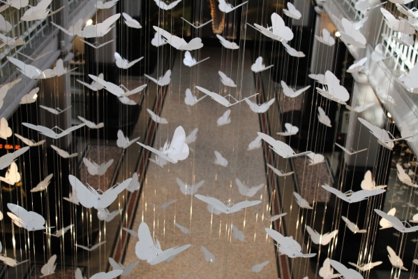
Butterfly mobile taken during my visit to Manchester Museum

This last month I’ve been so busy with writing and sorting out my data that I haven’t had a chance to do anything with my blog. In the last week, however, I’ve visited two more museums to look at some Neolithic and Bronze Age teeth.

A few days ago I took my first trip up to Manchester. I’ve never been to this part of the country but needed to go and visit Lancaster Maritime Museum and Manchester Museum. Both museums were lovely but I only really got a chance to have a look around Manchester Museum. It reminded me of a small Natural History Museum as it had quite a lot of animal specimens – including a lot of skulls! I took plenty of pictures of these and I might write some posts about them, like my old ‘skull of the month‘ posts.

It was really nice to go up north for a bit. I traveled up with my boyfriend, and it was lovely having the company! We also stayed with some friends who I haven’t seen for a couple of years. It was so nice seeing them! We even talked about going on holiday to the Lakes one day – something I’m very keen to do. Oh and they also had very cute cat called Arthur. He was such a sweetie and playing with him made me want one of my own. Maybe one day.

In other news the conference is going really well. I haven’t talked about it for a while but remember that I’m on the committee for a conference called Skeletons, Stories and Social Bodies? Well that’s happening at the end of this month. I can’t believe it’s coming up so soon, but everything is coming together and we have lots of people registered. I think it’s going to be really good, especially for a brand new conference! It’s really exciting and I’m really pleased to be a part of it. But before then I have two more collections to visit, and quite a bit of data analysis and write up to finish!

Return to Cardiff Museum

Sorry I didn’t post anything late week I was rather busy so it slipped my mind, so this post will talk about my last 2 weeks of work. This includes another trip to the stores of the Corinium Museum, volunteering at the Royal College of Surgeons, a trip to the Cotswold Archaeology office and another up to the National Museum of Wales in Cardiff.
My visit to the Corinium Museum stores was successful again. I got through another load of Anglo-Saxon skeletons for my data collection. It’s such a good collection and its going to be very useful for my PhD project as it includes lots of juvenile individuals. This makes me very happy! However, I’ve got quite a few more trips to make to their stores as it’s such a large collection.
The day after my trip to Cirencester I was back at the Royal College  of Surgeons volunteering in the museums department. It’s been a few weeks since  I’ve been there as the museum has been quite busy and haven’t been able to have me in. However, it was great to be back packing more skeletons into boxes ready for the move. Of course, it was also great to see the staff members again. I do enjoy working there!
So that was last week. I started this week by coming up to Kemble, which is near Cirencester. The purpose of this trip was to visit one of the office of Cotswold Archaeology, as they had a couple of Bronze Age and Iron Age skeletons. Whilst there I got to meet a couple of lovely people, including Sharon Clough who gave me some great information regarding some of the other collections I’m intending to use in my research. I also got to chat to Sharon about commercial archaeology units and learn a bit more about them. I’ve only really visited museums and universities so far, so it was a really good opportunity to learn about the commercial sector – a completely new area for me!
This week has ended with a trip but to the National Museum of Wales, in Cardiff. In my last visit I went through the prehistoric human remains in their collection. This time I was going back to the relevant specimens and taking measurements. I’ve now managed to get collection of Neolithic individuals recorded, plus a few Bronze Age remains, which is always good!
So another day, another lot of data collection completed. I think it’s all going well – I feel like I’ve got a lot done, but then I still have a load more to do! As a little fish called Dory once said ‘just keep swimming!’

Update from Me

20150612_183947So I’ve been pretty poor at maintaining this blog recently for a number of reasons. Over the last month or so a lot a has happened which has meant that I haven’t had the time or energy to keep up the writing. However, these events have been well deserved and long-needed (even if I do say so myself!). The first major thing was that my boyfriend and I finally got to move into our own flat, it’s only taken us 7 and a half years! Since moving in and buying nice things to furnish it with we are both so much happier. It’s amazing how much a difference having our own space in a nice part of town, with only the two of us to worry about. It’s been quite a long wait but it was totally worth it!

The second major thing that has happened to me recently is that I applied for a studentship at the Univeristy of Southampton to do a PhD in Archaeology. After I completed my masters a couple of years ago I wasn’t convinced that further study was right for me and so I took my current job at Reading Univeristy as a Research Assistant. This job has been amazing allowing me to stay in an academic environment and work with some great people, assiting their research. Over the past year or so I have been thinking about my future and career and struggled to find something that I wanted to pursue. I was then shown the job advert for the PhD Studetship and it sounded ideal. My reasons for not pursuing a PhD sooner revolved around the topic and cost, however this one ticked all of the boxes. The studentship aims to improve aging techniques for human skeletal remains in archaeological assemblages which could provide a positive contribution to the field – something that was important to me, plus there was the added benefit for being funded.

I am extremely pleased to say that this week I recieved confirmation that I had been awarded the studentship! I will be starting sometime at the end of September and I’m very much looking forward to it. It will allow me to work in an area that I am passionate, carry out my own research and to potentially meet a lot of interesting and exciting people. To be honest I can’t really believe it still but I’m sure it’ll sink in at some point!

Getting to this point has not been easy – for myself or my family. I have very nearly given up on pursuing a career in the anthropology/archaeology field on multiple occasions even though I knew that wouldn’t make me happy. I have always heard, and even said myself, that you should do something that makes you happy but that it so much easier said then done. It is really difficult pursuing your dream job, especially if it’s in a slightly niche subject or if you need lots of work experience to get anywhere, and getting a suitable income to provide for yourself. I am hopeful in saying that I think that my PhD is that start of my career in a subject I really enjoy, but it has been sheer determination and a lot of support from my family and boyfriend that has really got me through. I feel very lucky to have gotten here, and yes I have worked very hard to get here, but I still feel lucky.

If you are trying to pursure your ideal career, or are attempting to get into a difficult field – keep going. Work hard, be nice to people and take any opportunities that you can manage – you probably won’t be able to do everything but showing you tried will count. Also make sure you are surrounded by people who support you and who you can depend on. If you’re going through things like I have over the past year or so you’ll need help and someone to turn to when you are feeling bad about yourself and your decisions. They are invaluable and are honestly the reason why I have managed to get this far.  However, finally remember that there is an element of luck in all of this. I was luckly to see the job/PhD advert when I did, but that doesn’t mean it won’t happen just that it might happen when you don’t expect it!

I don’t usually write posts like this, and to be honest I didn’t really intend to when I first sat down today, but I felt like I’ve managed to get some things off my chest. I also hope that if anyone who has been in a similar position to me over the last year reads this I hope this post can bring them a little comfort or advice. You’re not alone, and keep your head up – I’m pretty certain it will work out in the end!
Week 35 Volunteering at the Royal College of Suregons

Outside of the Royal College of Surgeons. Image taken from http://nobelbiocare-eyearcourse.com/fgdp.html.

Outside of the Royal College of Surgeons. Image taken from http://nobelbiocare-eyearcourse.com/fgdp.html.

So have a nice chilled out Christmas break I’m back at the college looking at more interesting specimens. As this is my forst week back, including my job at Reading University, I am still getting adjusted with actually having to work as having two weeks off at Christmas was great but as I did very little it’s taken me a few days to get back into the swing of things.

This week at the college I had quite a few skulls again with similar conditions to those I’ve examined in the last few weeks. However, I also had two partial skeletons and one complete skeleton. All three specimens had the same condition of iniencephaly, which I discussed in week 33. This condition is a result of a neural tube defect and therefore causes poor development of the skull, vertebrae and ribs. The two partial skeletons that I had only included these elements in addition to the pelvis so it was really interesting to compare them to a complete skeleton with the condition. It was clear to see that all of the other bones had developed as normal, although the scapulas were displaced as a result of the curvature of the spine. It was an extremely delicate but facinating specimen and I very much enjoyed working with it.

In addition to my work with the bones I was also shown a 3D-printed model of an infants skull. The conservation team had recieved some funding to help produce teaching models for the surgeons who use the Wellcome Museum of Anatomy and Pathology. One of the lecterures had requested to get an infant skull that they could handle as it would be very useful for teaching purposes. It was possible to provide them with real infant skulls as they are so delicate and easily damaged and therefore the idea came to try 3D printing. The model that was produced was amazing and fairly detailed – however, there was a slight problem with it being far too small. It means that it can’t be used for teaching as it is not accurate but it does give hope that this method could be used in the future.

Week 34 Volunteering at the Royal College of Surgeons

I was meant to post this a week ago but times got away with me given its Christmas and all! Any way here is my post about last week’s volunteering at the Royal College of Surgeons.

Today was my last day volunteering before I break for Christmas. I must admit finishing on a Thursday is really nice – not only does it make it a short week but I get to finish on my bone work. This makes me happy.

I had a few interesting specimens this week including a few with the condition I discussed last week, iniencephaly. These specimens are amazing to look at – particularly if you’re an anthropologist like me!

In addition to the skulls I had some legs to look at too. These were mounted in perspex boxes and were incredibly small. Half of these specimens appeared to be casts of the other legs which had been modeled to show the epiphyses and patellas. This was concluded after I realised that the pairs of bones had identical labels with the same information, including the original creators specimen numbers.

I’m him to enjoy my break now over Christmas and I’m very fortunate to have so much time off (the university where I work closes over Christmas!). In the mean time I wish everyone a Merry Christmas and a happy new year!

image

Christmas at the Royal College of Surgeons

Week 32 Volunteering at the Royal College of Surgeons

Outside of the Royal College of Surgeons. Image taken from http://nobelbiocare-eyearcourse.com/fgdp.html.

Outside of the Royal College of Surgeons. Image taken from http://nobelbiocare-eyearcourse.com/fgdp.html.

This week I was in on Tuesday instead of Thursday because there were the volunteer Christmas drinks in the evening. It was really nice and interesting to meet some of the other volunteers as well as spending time with the museum staff outside of the normal hours. The director made a nice speech thanking all of the volunteers and I even received a box of chocolates from the curator of the Wellcome Museum of Anatomy and Pathology as a thank you, which I thought was very sweet!

My boxes of skulls took me a little longer to go through today as many of them were not articulated/glued together. This meant that I had to check which cranial elements were present and take a photograph of each one. Although this more of a lengthy process it definitely tested my knowledge of the developing skull. I have handled many adult skulls during my studies and time at the College but very few infant skulls. They are extremely delicate and fragile so I have to be very careful when handling the specimens. 

Image of the temporal bone at birth, depicting the tympanic ring. Image taken from: http://en.wikipedia.org/wiki/Temporal_bone

Image of the temporal bone at birth, depicting the tympanic ring. Image taken from: http://en.wikipedia.org/wiki/Temporal_bone

These specimens also gave me a chance to see the tympanic ring which forms part of the temporal bone (see fig. 1). I also saw many deciduous teeth that had fallen out of one of the mandibles. I’ve seen many teeth in the jaw but very few then they have fallen out. They are amazingly small but it is still possible to identify which teeth are present because of their distinctive shape. However, the best  thing that I saw was one of the ear bones which had been isolated. I have only ever seen pictures of the ear bones so it was amazing seeing this one from an infant specimen, the particular bone that was present in this specimen was the incus. There are three ear bones: the incus, malleus and the stapes. Their colloquial names are the anvil, hammer and stirrup due to their shape. These bones are so tiny that it is easy to see why few ear bones are recovered from archaeological digs. It is times like these that make me realise how fortunate I am with this position. I wonder if I’ll come across any more ear bones in the remaining boxes I have to go through!

The incus bone, one of the three ear bones. Image taken from: http://wellnessadvocate.com/?dgl=10257

The incus bone, one of the three ear bones. Image taken from: http://wellnessadvocate.com/?dgl=10257

The identification of Vitamin D Deficiency in Adults from Post-Medieval Sites.

Image of pseudofractures occurring at the scapula spinous process. Image taken from Ives & Brickley (2014) p. 49.

Image of pseudofractures occurring at the scapula spinous process. Image taken from Ives & Brickley (2014) p. 49.

I haven’t been around to do a post on a particular pathology for a while but as I was looking through the International Journal of Paleopathology I came across an article looking at osteomalacia. During my pathology course at Uni we touched on this and therefore thought it would be a good article to focus on.

Osteomalacia is a softening of the bones due to a vitamin D deficiency. This is commonly known as rickets in children. It is often identified in juveniles by the characteristic bowing of the long bones. As vitamin D is used for enhancing the intake of calcium and phosphorus it is vital for solid and strong bone growth. A reduced level in these minerals delay and prevent the mineralization of newly formed organic bone matrix resulting in bowing and a weakening of the bones.

Although osteomalacia can be fairly straight forward to identify in children it is much harder in adults. As the long bones have fully developed in adulthood no bowing occurs but a period of vitamin D deficiency can cause a weakening of the bones producing pseudo fractures. These differ from fractures caused by accident and injury as small linear or stress fractures are produced at weakened sites. In Fig. 2 a microscopic view of osteomalacia is presented with arrows highlighting areas where mineralisation has not occurred.

There are two ways in which vitamin D is obtained. The first has been briefly mentioned and occurs by being exposed to UV radiation. The second is by consuming particular food including oily fish and eggs. In modern life these things are easy to come by and therefore the prevalence of osteomalacia is low. However, in the post-medieval period, which the study sample is from, people worked in dark conditions will little exposure to the sun and oily fish or eggs were hard to come by as it was expensive. This resulted in a higher rate of vitamin D deficiency and Osteomalacia during this time.

This article by Ives & Brickley (2014) investigated the skeletal manifestations of Osteomalacia in a large sample dating to the post-medieval period. This includes examining 1181 individuals from 6 post-medieval urban sites plus a collection of 142 burials from a previous study (Brickley et al. 2007) from Birmingham. This gave a total sample of 1323 individuals to examine for the presence of osteomalacia.

Microscopic image of osteomalacia. Image taken from Ives & Brickley (2014) p.51.

Microscopic image of osteomalacia. Image taken from Ives & Brickley (2014) p.51.

When examining the skeletons the spinious process of the scapula appeared to be a common site for pseudo fractures. In total there were 9 cases of this type of fracture, of which 6 also had pseudo fractures in the ribs. Other sites of these fractures also included the pelvic girdle and vertebrae (see fig. 3 for all off the locations of pseudo fractures from this study). Microscopic analysis of some individuals also showed a weakening of the bone and were comparable to the study by Brickley et al. (2007). This method of detection was also used on individuals who did not have any pseudo fractures of the scapula spinious process, but in the femoral head and ribs to confirm the diagnosis of osteomalacia.

From the sample 19 out 1323 individuals were identified as having osteomalacia giving a prevalence rate of 1.43%. When this result is compared to what is known about rickets in children of this period this rate is very low. The authors of this paper provide a few explanations for this. A conservative approach was taken with the study assigning osteomalacia only to those individuals with strong indications of the condition. They also suggest that some adults may have suffered from vitamin D deficiency for a short period and therefore had fully recovered which meant the traits of the condition were not viable on the skeleton. Finally the authors state that between pseudo fractures can be difficult to differentiate from those produced from injury and as they were taking a conservative approach the individual would not have been included.

From this study it appears that you osteomalacia can be accurately identified using characteristic patterns of fractures. It was found that at least 11 individuals from the sample had died before acquiring adequate levels of vitamin D, however in some cases a skeleton may have been so fragile from the deficiency which would complicate an accurate diagnosis. The findings from the study also suggest that individuals who lived in the post-medieval period may have gone through multiple episodes of vitamin D deficiency though these may not have been severe enough to cause a collapse in the bone.

The identification of multiple sites of the skeleton where pseudo-fractures formed provides new information about osteomalacia, and the microscopic analysis confirms this. Although these fractures may not always be present they may be used an indication for the condition and easily appilcable to future study samples. 

The skeletal locations affected by pseudofractures together with the true prevalence rate or crude prevalence rate of fracture occurrence. Image taken from Ives & Brickley (2014) p.52.

The skeletal locations affected by pseudofractures together with the true prevalence rate or crude prevalence rate of fracture occurrence. Image taken from Ives & Brickley (2014) p.52.

References.

M. Brickley, S. Mays, R. Ives (2007).l “An investigation of skeletal indicators of vitamin D deficiency in adults: effective markers for interpreting past living conditions and pollution levels in Eighteenth and Nineteenth Century Birmingham, England.” Am. J. Phys. Anthropol., 132: 67–79

Ives, R. and M. Brickley (2014). “New findings in the identification of adult vitamin D deficiency osteomalacia: Results from a large-scale study.” International Journal of Paleopathology 7(0): 45-56.

Week 23 Volunteering at the Royal College of London

Outside of the Royal College of Surgeons. Image taken from http://nobelbiocare-eyearcourse.com/fgdp.html.

Outside of the Royal College of Surgeons. Image taken from http://nobelbiocare-eyearcourse.com/fgdp.html.

Today I got to start on the new project at the college and pretty pleased I can do it!

I think I’ve mentioned what this project invokes but I certainly don’t mind explaining it again! This project is similar to the last one as I am having to record and inventory another collection. However, the new project involves a particular collection at the museum has. It is quite a small collection, I think there are about 160 objects, but it’s very interesting as well as very sensitive. This is because it includes individual who most likely died before birth because of a pathology.

A few years ago a couple of students came in and went through this collection and made notes including any pathology observed. As the curator was not involved in this project she has asked me to go through the boxes and check their contents see if the students descriptions/comments are correct.

This project is going to test my knowledge in pathologies as well as perinatal remains. I’ve very excited about this project and am very much looking forward to getting stuck into it!

Week 22 Volunteering at the Royal College of Surgeons

Outside of the Royal College of Surgeons. Image taken from http://nobelbiocare-eyearcourse.com/fgdp.html.

Outside of the Royal College of Surgeons. Image taken from http://nobelbiocare-eyearcourse.com/fgdp.html.

Today I wasn’t feeling great- I’m not sure what’s up I just didn’t feel like myself however the thought if going in to London made me feel a lot better. I also wanted to be in today as I knew that it would be my last week on the current project before moving on to the new one.

It was a great week to finish this project on as I had some great specimens to record. However before I got into the interesting bits I had to finish off from where I left last week. This meant going through the last few boxes of foot bones. These included the navicular, cuboid and the cuneiforms and was therefore tested again on my ability to side these bones. I think I did quite well as I had the Tim White Human Osteology book next to me, which is always useful!

After the foot bones I moved onto two boxes containing perinatal bones. These are bones which belong to individuals who died either at birth or around that time. When you stop and think it is very sad however these specimens are extremely useful to teaching collections. I have also become slightly desensitised to their context and instead I am fascinated by their size and shape, as the bones are very to their adult size.

The first box I looked at had multiple perinatal skulls as well as vertebral column. These were all articulated and it was amazing being able to hold these specimens as it is rare, particularly in archaeology, to get complete infant bones. The bones were so fragile and light that careful handling is needed and some of he specimens had been previously damaged. I have handled enough bones in my university career to be confident when doing this and therefore had no problems.

The final box of the day had a complete perinatal skeleton which was incredible and an amazing thing to finish the project on. As I have already said it is rare to find complete skeletons in archaeological excavations and therefore I don’t think that I’ve ever seen a complete infant skeleton at first hand. It is amazing how many bones there are and how tiny they are! None of the bones of the vertebrae were fused so they were in 3 parts and to get an idea of the size if this individual the femur was the same length as the back of my hand.

I really love looking at these type of skeletons and I’m not sure why. I just find them fascinating and I wish I had the chance to see more. However, hear collections are rare as they are small in number. For example the specimens that I recorded today were the entire handling collection. There are some others in the stores but they will be either too fragile or valuable to be given to eager students! Another reason why I enjoy this type of work is that I can attempt to age the individuals and by doing this use the information I learnt at University and also learn some new things. For example as I have had to age individuals that were so young before I had to use a different method. This included measuring various points and lengths of the bones to get good age estimate.

I really enjoyed today and it was just what I needed after feeling a bit under the weather this morning. It was a very good day and excellent specimens to finish this project of creating a bone inventory. I am now looking forward to coming back next week and starting on the project which will be involving even more perinatal skulls! Bring it on!

Week 21 Volunteering at the Royal College of Surgeons

Outside of the Royal College of Surgeons. Image taken from http://nobelbiocare-eyearcourse.com/fgdp.html.

Outside of the Royal College of Surgeons. Image taken from http://nobelbiocare-eyearcourse.com/fgdp.html.

So after a week off and away in sunny Portugal I was back at the college to sort through the remaining boxes of bones. I am very close to finishing the current project of creating a bone inventory and hopefully next week will be the last week sorting out boxes of bones. I’ll then move on to infant skulls which have various forms of pathology.

This week, however, focused on the lower leg. To start with I had two boxes of articulated lower legs which I got out and divided into lefts and rights. These we’re very simple to organise and took me very little time to sort. There weren’t any pathologies but it was nice to see how the bones fitted together and articulated with one another. I think it is these specimens which may be particularly useful for the students as they can get a good look at how the bones move.

I then had some individual foot bones to sort and included the metatarsals and foot phalanges. I had to count out the individual bones and make sure that they were in the correct boxes. I suspect that some of the elements had been mixed up by previous students whilst studying. I can see how this can be done but it doesn’t make it easy for any future individual to look at the bones properly.

After these small bones I was presented with the ankle bones. I only got through the talus and calcaneus bones before stopping so I have the rest of the ankle to finish next week. I stopped early because I wanted to have a look at the wet lab where wet specimens are restored and conserved. I was talked through some of the processes and techniques which are used and why various things had to be done. There were some very interesting specimens including an elephants foot and a raccoons skull. The conserver’s job is really interesting and every day is different. There is always something to repot, refill or replace and the specimens range from skeletal to soft tissue, animal to human and large to small. Hopefully I’ll get to see even more in the future!