Home » Posts tagged 'teeth'
Tag Archives: teeth
A Night In An Old Operating Theatre!

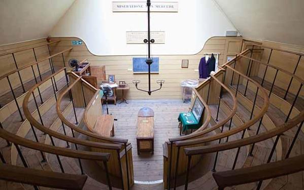
The Old Operating Theatre. Image taken from http://www.telegraph.co.uk/travel/destinations/europe/united-kingdom/england/london/articles/London-in-your-lunch-break-the-Old-Operating-Theatre/
This week has been a long one! I’m not sure why as it’s been pretty good and quite productive but it’s taken a while to get through. Maybe it’s because I’ve been travelling for my data collection again and I’m not used to driving so much?! As well as my PhD work this week I went to a really cool talk about Bodysnatching in an old operating theatre – perfect for Halloween!
On Monday I was back at the stores of the Hampshire Cultural Trust to finish going through the various sites they have. I’m pretty pleased with myself as I’ve managed to get through a lot of skeletons in a decent amount of time. There are two small sites to work through but as they’ll only take me half a day at most I will return another time. At some point in the future I will need to go to their other store to access a Romano-British population.

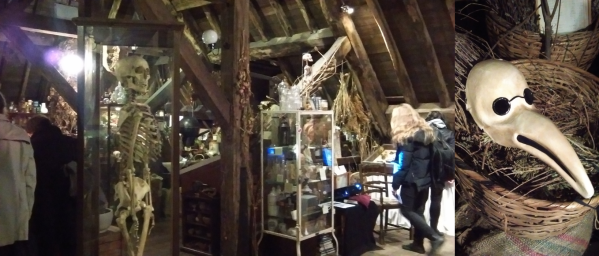
A view of the museum at the Old Operating Theatre, and a replica beak mask.
Data Collection in the Cotswolds

My last blog post found me in Cardiff to visit the National Museum of Wales, to see their human remains collection. Since then I have continued with my museum trips and data collection, and so far so good!
On Monday I went to the Museum in the Park. A local museum in Stroud, a town in Gloucestershire. Although my boyfriend lived there when we first went out I never got round to visiting the museum, so here was a great opportunity. It’s a lovely museum located in a beautiful park so is a great place to visit with the family. Unfortunately I didn’t get a chance to go round the museum apart from walking to a few display cases to measure a couple of skulls! However, from what I did see it looked really nice and well laid ou t- certainly a place to go back and visit.
Whilst at Museum in the Park I was able to measure a number of teeth dating to the Neolithic for my PhD research. These predominately consisted of mandibles but as Neolithic material isn’t great in number these are a welcome addition! It was great working with the collection and I have to say a special thanks to the Documentation and Collections Officer for the museum, Alexia Clark. Alexia was extremely helpful and accommodating and I very much appreciated her help. I don’t see myself heading back to Stroud Museum to collect any more data but if I’m in the area again I may make a special trip to have a proper look around.
In addition to Stroud I also went to the stores of Corinium Museum, Cirencester. As I was born in Swindon, about 20 miles away, I went to the Corinium Museum as a kid. However, I only really remember the Roman exhibits and displays that they have. For this trip I was again looking at Neolithic remains from the site of Hazleton North. Again, I managed to examine some lovely Neolithic teeth, there was also the added bonus of a complete individual and a number of skulls. This is pretty impressive as many of the Neolithic material is dis-articulated and therefore it is difficult to determine specific individuals. This collection will be a great addition to my research.
At some point in the future I will be returning to the Cirencester stores as they have at least one other collection that I wish to use. This is the Anglo-Saxon material from Butler’s Field. Plus there may be a few additional sites dating to the Bronze Age and Iron Age, so I will definitely be going there again soon. Again, the staff at the museum have been incredibly helpful and so everyone I have met have been amazing. They definitely adding to my PhD experience and reinforces my desire to work within the museum sector in some capacity one day!
Although my data collecting has so far been straightforward and without any issues there is one aspect that leaves food for thought. Whilst at working through the Hazleton North material I found that a number of teeth, predominately molars, had been removed for isotopic analysis. This of course means that I cannot use them for my project. This isotopic work has increased the understanding of the individuals within the collection, including what food they ate and where they originated. In some aspects this will aid my research as the diet can be determined, which is vital for understanding the factors contributing to dental wear. On the other hand, I am now unable to include those teeth in my own research. This means that there are some individuals that I can no longer use, as no molars are present, therefore reducing my sample size. I see this an unavoidable annoyance. I respect the other researchers, and certainly their research will contribute to my own work in an alternative way, and most importantly their work will provide useful insights of the past. None-the-less, I can’t help but feel a twinge of irritation – especially if it effects a juvenile individual!
Next week I hope to visit some of the collections held by Hampshire Cultural Trust, and in the mean time I have to finish taking measurement from my photos of the collections and attend a friend’s engagement party, oh and play two hockey matches! At some point I will have a day off!
New Page – Identifying Molars
It’s been a while since my last post and I’ve been meaning to create a page for identifying and distinguishing molars a while now, but I’ve finally gotten round to it.
At university and during my time as an undergraduate I found it quite difficult to distinguish between the different teeth – particularly the molars. As my PhD project focuses on these teeth I had to quickly gets to grips with identifying molar teeth correctly. I’ve therefore created a new page to help other osteologists out there who need some extra help!
This page only includes the upper and lower permanent molars as they are the teeth I am most familiar with. Also, some of the tips and features I have mentioned below are from my own observations although the majority come from Simon Hillson’s book ‘Dental Anthropology,’ (1996) which I highly recommend if you are going to spend any time looking at teeth.
Go and check it out here! Also, I’m always happy to receive feedback and comments 🙂
Week 46 Volunteering at the Royal College of Surgeons

Outside of the Royal College of Surgeons. Image taken from http://nobelbiocare-eyearcourse.com/fgdp.html.
I’ve been a bit quiet in this blog recently as I have moved house so there were of things to sort out . However , I have still been volunteering and gave made great progress on the Stack Collection of deciduous teeth.
I have now finished photographing and recording the specimens and I can honestly say my ability to identify teeth has greatly improved. Teeth were not always a strong point for me, mainly because we did not have a lot of practical experience with them at university. However, by using the common osteological text books (including Simon Hillson’s Dental Anthropology, Tim White’s Human Osteology and ‘The Skeleton Keys’ by Schwartz) and drawing on my own memory I was able to identify the teeth quite quickly. By the time I came to the of the specimens I felt reassured of my ability and therefore went back to the first specimen to check them. This was a good idea as I did make some corrections and was able to clarify some of the teeth I was unsure about.
As I have finished with the actual specimens I have moved on to recording the information written in a catalogue of dentition, which was created by the collector. This is enabling me to check some of the information I have already obtained from associated cards as well as adding comments from the Pathologist’s Report. So far these have include cardiac defects, defects associated with the Central Nervous System and pre-eclampsia. I recognised some of the conditions as they appeared in the previous project such as spina bifida and intra-cranial haemorrhage. There were a few defects I did not recognise but a quick Internet search revealed the condition.
I will not be in next week as I am switching my work days round so I can attend the proffered papers meeting for the Society for the Study of Human Biology. I am very much looking forward to going to this meeting as there are going to be some very interesting papers being presented. This includes one by Dr Liversidge who has previously used the collection I am currently working so I intend to introduce myself to her.
Week 42 Volunteering At the Royal College of Surgeons

Outside of the Royal College of Surgeons. Image taken from http://nobelbiocare-eyearcourse.com/fgdp.html.
This week at the College I started photographing and recording the deciduous teeth in the current collection. This is an extremely delicate task as the teeth are so small and fragile. I had to carefully line the teeth up, arranged by tooth type, and take a photograph using a 1cm scale bar for reference.
It has been a little while since organising and arranging teeth so I had to refer to some textbooks to be certain. I attempted to arrange the teeth by type (incisor, canine and molar) and where possible I identified whether the teeth came from the maxilla (upper jaw) or mandible (lower jaw). This wasn’t too difficult for the incisors but I couldn’t always identify the molars and it was very hard to work out which jaw the canines came from. The difficulty of identifying theses teeth was a result of their size and age. The individuals I was working with were fetal or neo-natal and therefore only a small amount of dental development had occurred. This means that there is little to no development of the roots resulting in the crowns of the teeth being present, for example the canines only consist of a small triangle of enamel. The image below in the first pictures gives you an idea of the stage of development I am dealing with.

The stages of deciduous tooth development. Image taken from here
By the end of this project I am going to be very well experienced in handling, tiny specimens as well as increasing my knowledge of deciduous teeth.
Week 40 at the Royal College of Surgeons

Outside of the Royal College of Surgeons. Image taken from http://nobelbiocare-eyearcourse.com/fgdp.html.
I had a shorter day at the College this week because last Saturday I was hit in the head with a hockey ball so at the moment I’m a little prone to small headaches. I had a very impressive black eye, which I’ve never had before, that has gone a wonderful shade of various colours! However, it looks a lot worse that in was, the most annoying thing was that I was hit about 5 minutes into the game. Anyway I had another good day at the College, black eye and all!
This week I carried on with the digitalisation of the cards associated with the collection I am working with at the moment. These cards are for each set of deciduous teeth that are in the collection and include information about the owner of the teeth. This is very sensitive data and some even have the pathologies that the individual had. I’m therefore learning even more medical terms and conditions which is very interesting, there are even a few that I have recognised from medial drama such as Grey’s Anatomy (I’m a late comer to the show but I’m totally hooked! Thankfully I have Amazon Prime and watch multiple episodes at a time).
Next week I might take a break from the cards and start on the teeth.
Week 39 at the Royal College of Surgeons

Outside of the Royal College of Surgeons. Image taken from http://nobelbiocare-eyearcourse.com/fgdp.html.
After a week off due to moving house I’m back at the College where I’m starting on a new project. This time I will be working with a collection of infant teeth.
As with the previous protects I’m creating an inventory of the collection by taking photographs of the specimens and the associated paperwork. This information will then be uploaded and added to the museums database.
The collection includes the teeth of fetal or neonatal individuals. Each set of teeth have some personal information associated with them making it a very sensitive collection. I’m not sure why such a collection war created but it has a very high potential for research due to the quality of the specimens and it’s data.
When I first started my course looking at human remains at university I was never a huge fan of teeth. However, as time has gone on I’ve become more familiar with teeth and their uses in research. If you know how to use them teeth can tell you a lot including the age and diet of an individual. During this project I hope to learn even more about teeth and what information they can provide.
A Look at the Accuracy of Dental Age Estimation Charts
So it’s now the 4th of July and I haven’t created a new skull of the month and I don’t think I will. Over the past few months I’ve been really rubbish at updating my blog and the reason behind that – I’ve been applying for jobs and working out what I’m going to do at the end of July (my current contract is ending and I have to decide whether to re-sign it or not!)
Anyway, because of all this I’ve decided that for July I’m going to try and catch-up with some of the articles that I’ve wanted to share over the past couple of months. Some relate to various skulls of the month whilst others are just articles of interest. Ideally, I’m going to do a couple a week (depending on how many applications I have outstanding!) so fingers crossed!
To start with I’m going to read an article by AlQahtani, Hector and Liversidge called ‘Accuracy of dental age estimation charts: Schour and Massler, Ubelaker and the London Atlas’. During my osteology courses at university I used both the Schour and Massler and Ubelaker for estimating age from dental remains. I came across the article a while back and I just haven’t got round to reading it.
This particular study caught my eye because it’s aim was to compare the accuracy of estimating age from developing teeth from the above methods. When using these types of methods, using illustrations and charts to form an estimate, it is important that they are reassessed over time to test their accuracy. This is essential in subjects like archaeology and anthropology where the outcome of the result could be very fundamental.
I have discussed in the past how teeth are used for aging skeletal collections. These answers help to provide a profile of the individual and in forensic environments could potentially lead to the identification of a missing person. In archaeology age at death is used as an indicator of a populations health, providing an insight into past life and communities. In both situations obtaining an accurate and reliable age estimate is key, and therefore it is necessary that the common methods used for this process are assessed periodically.
The article by AlQahtani, Hector and Liversidge first provides a small amount of background to each of the chosen methods under study. This image of Schour & Massler should be familiar to any osteology student. It depicts 21 drawings of dental development from the age of 31 weeks in utero up until adulthood. The method was apparently criticized in the early days due to the lack of information about the material or the method to provide the images. Since the original production in 1941 revisions have been made of Schour & Massler’s atlas with the use of radiographs.

Schour & Massler’s 1944 chart, taken from A Test of Ubelaker’s Method of Estimating Subadult Age from the Dentition (E. Smith 1999) p.30.
In 1978 Ubelaker attempted to improve the method for obtaining age estimates from dental development and used a number of published sources. This method was seen as one which covered a the range of variation that can been seen at each stage of development. Again, most osteologists will have seen the following chart.

Ubelaker’s 1989 chart, taken from A Test of Ubelaker’s Method of Estimating Subadult Age from the Dentition (E. Smith 1999) p.31.
Finally, there is the London Atlas which has combined many resources to create a dental chart that includes 31 age categories. This cart is tooth specific and defines each tooth by the development of it’s enamel, dentin, and pulp cavity. The other bonus is that this chart has been made freely available here.

Second page of the London Atlas showing the development of individual teeth available here.
That’s the method under investigation briefly covered now onto the study. As I have already mentioned the aim of the study was to assess the accuracy of estimating age from developing teeth using the three methods above. In order to do this a large sample of skeletal remains of known age at death was used. This included 183 individuals who were aged between 31 weeks in utero to 4.27 years and a further 1323 individuals (649 male and 674 female) aged between 2.07 and 23.86 years. In addition to skeletal remains the archived dental radiographs of living patients were also used. From this the age estimates were made using the three methods chosen for study and compared to the chronological age. To determine the reliability of the aging methods statistical analysis, including a paired t-test was carried out.
Before moving onto the results it is worth mentioning that intra-observer error was taken into account. This means that the same individual contacted the age estimates multiple times of a sub-set of the study sample. From doing this it was found that the error rate was low, meaning taht there was excellent when repeated for all three of the methods.
So the final results. The method which produced the closest age estimate to the chronological age was the London Atlas. For the other two methods it was found that they both under-estimated by around 0.75 years. This result was attributed to the amount of categories produced by Schour & Massler and Ublekaer as their age categories are much larger as the individual ages. This potentially misses out important development phases which can provide a more accurate age estimate. This is true of the growth of the third molar. As can be seen in the dental charts above the development of this molar is not depicted in detail, unlike the London Atlas. Due to this AlQahtani et al. removed the age estimates using the third molar and discovered that both the Schour & Massler and Ubelaker charts still under-estimate the age but only by 0.5 years. In comparison the London Atlas had a mean difference of 0 years.
This study showed, that whilst all three methods of estimating age using dental development is fairly accurate the London Atlas performed the best. As stated above these tests are important to the study of osteology and anthropology. This is because determining the age at death is vital of identifying individuals and establishing the health of a population for their dead. These studies cause researchers to constantly check their methods and to further their work in order to obtain the most reliable system that is possible, which is extremely important.
References
S. J. AlQahtani, M. P. Hector and H. M. Liversidge (2014). ‘Accuracy of dental age estimation charts: Schour and Massler, Ubelaker and the London Atlas.’ In the American Journal of Physical Anthropology Volume 154, Issue 1, pages 70–78, May 2014
E. SMith (1999). ‘A Test of Ubelaker’s Method of Estimating Subadult Age from the Dentition’. A Thesis Submitted in Partial Fulfillment of the Requirements for the Degree of Master of Science in Human Biology in the Graduate School of the University of Indianapolis May 2005.
BBC Article – The Narwhal’s Tusk is a Sensitive One
I was just looking through the various news stories on the BBC and came across this one by Ella Davies. A resent study at Harvard School of Dental Medicine has found that a Narwhal’s tusk is sensitive to temperature and chemical differences to the surrounding environment. This was established when a difference in the heart rate of a Narwhal occured when exposed to these changes.
The Narwhal is a pretty cool and interesting creature. The tusk is usually found on males and actually a front tooth which grows to extrordinary lengths, up to 2.6m (9ft). It has long been suggested that the tusk is a sexual characteristic, as it only usually males who have them. However, this new study suggests a new insight into the Narwhal – although more reserach is needed in the area to establish what is actualy going on. As these animals are rarely sighted very little is known about them in any sense so any new information is extremely important and revealing. The research is now continuing and information from hunters is being gained to attempt to shed more life on these elusive creatures.
Read the full article here.

