My British Osteological Database – An Update

skele_1

Finding research material is major component for any PhD. This can often mean extensive online searchers, emails to curators and searching through journal articles for relevant or useful material.

From my own experiences (where I needed large samples of human molars from a range of archaeological periods) this is not always an easy thing to do. A while back I decided to produce a crude database about some of the human remains collections I came across during my search. Recently, I decided to update this to make it more user friendly and to add a couple more sites.

You can find a downloadable excel file here with site names, dates and basic collection information.

Enjoy!

PS. If there are any more collections that you know of (and their information can be shared publicly) please get in touch!

Where has Christmas gone?!

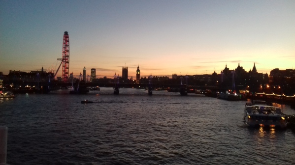
20170119_165742

A shot of London from Waterloo Bridge, taken on my way home from the Hunterian Museum.

My last blog post was before Christmas so that’s nearly a month ago now. How time flies when you’ve got work to do! I would say my new years resolution would be to write more blog posts, but I’m not sure if I’ll have the time. As well my PhD work, the conference I’m a committee member for is getting ever closer! Maybe after March (and the conference) I’ll have some more time. I really want to expand these blog posts to more than ‘what I’ve been up to’ – maybe I’ll have the time soon!

Ah, Christmas feels so long ago now, but it was lovely. I managed to have a week off to see family, catch up with some old friends and generally chill out for a bit. It was great – even with a horrible cough and cold! But a week goes pretty fast when you’re having fun and I was soon home again and back carrying on with the PhD.

The PhD stuff is going well, I’ve booked to go and visit some new museums (that have Neolithic remains hooray!) and started going down to visit the Dorset County Museum. It’s pretty far to go but they have some really useful collections. Plus their stores are in a church, which is quite interesting if a little cold! I’ve said before that I really enjoy going to these museums as I get to meet new people. This week that included Claire Randall a zoo- and osteo-  archeologist. We had a lovely chat and it was great finding out about her work. This week I’ve also been brushing up on my statistics, which I have a love-hate relationship with! It can be a pain to get through but it’s so satisfying once you’ve done it and got it right!

Enough about the PhD work. The other cool thing I’m involved in at the moment is the Skeletons Stories and Social Bodies conference. I’ve talked about in many times (and you can find out even more by visiting our website) but it’s now getting quite close!  We’ve now had all of the abstracts in and produced a draft schedule. I am really looking forward to it as we have some great presentations covering a wide range of things! I know the speakers will be great but I also can’t wait to meet some of the delegates. There are quite a few people on social media who have been really supportive and enthusiastic about the conference and it would be great to finally put some names to faces! Not to long to wait now 😀

Finally, today I was back helping pack the collections at the Hunterian Museum. I think I asy this every time but I do enjoy working there. The people are great and I love the museum, but it also gives me some head space away from my PhD work. It’s so easy to become obsessed and constantly worried that I should be working that it’s nice to escape once a fortnight! So I’ll be back there in two weeks time, still packing away.


The New Plan – Weekly Updates!

So I’ve been a bit rubbish with my blog (again!) but Ive told myself that I am going to be better from now on. I guess I haven’t written much as I haven’t felt that there’s much to say. HOWEVER, I am now back volunteering at the Royal College of Surgeons and I’m going to start visiting museums to access their collections for my PhD work. That means there should be plenty to talk about so I have no excuse for not keeping up with the blog!
So first things first, I’m back at the Royal College of Surgeons volunteering. It’s been about a year since I was last here but I couldn’t help but come back! I enjoyed working here so much and the people are lovely so I was just waiting for the right time.
Now I’m back I’m doing something slightly different then previously, where I was tasked with creating inventories for a couple of collections. This time I’m packing objects/human remains ready for decanting and moving them to a new site. This is because the building in which the museums department is based is getting renovated and so most of the collections have to be moved off site. So far the boxes of remains I have been working on have been straight forward. This has included sorting a couple of boxes of loose ribs and one half (a complete right side) of a skeleton. I’m having to think about how best to wrap and pack each box so that the bones are safe and secure, ready to be moved. As I said the boxes so far have been relatively simple to sort out, but I’m sure there are going to be some tricky ones coming my way!
So that’s my volunteering stuff, now for a quick update about my PhD. I’m now at the stage where I am contacting museums in search of human remains that would be suitable for my project. At times this has been fairly straightforward. I’ve identified a collection, either from some literature or an online resource, then contacted the museum and have been able to find exactly what I am looking for. On other occasions it’s been a little more complicated. For example, I may have found the original excavation report that identifies a collection but it is then difficult to locate the remains. In these situations I have contacted the most likely institutions or commercial archaeological company and went from there. It’s taken a little bit of time but I now feel that I have identified a good amount of skeletal collections, at least as a starting point.
So what’s next? Well I’m visiting the National Museum of Wales in Cardiff and Cirencester museum over the next 2 weeks. Here I  hoping to have a preliminary look at the remains that they have to see how suitable they are for my project. I will (hopefully!) arrange another date to go back and measure any of the remains that can be used in my research. In the mean time I’m going to start contacting the museums that have collections that are appropriate for my work and get some dates for visits in the diary. My aim is to see and measure as many skeletons as possible before April next year, when my PhD upgrade will be. So lots of work to do but I can’t wait to get started with the data collection!
As I said before, now that I’ll be visiting new places and collections, plus the volunteering, I should be able to write an update of my progress and experiences every week! Here’s hoping! Skeleton-Hands-Facebook-Cover

New News!

In the last week two cool things have been confirmed:

I’m really excited about both of these and can’t wait to get stuck in.

The Volunteering

hunterian

Inside the Hunterian Museum. Image taken from here.

About 2 years ago I started volunteering at in the museums department of the Royal College of Surgeons and enjoyed it so much that continued to work there until I started my PhD. Whilst there I was lucky enough to work with some amazing osteology collections and saw some interesting pathologies. I really enjoyed working there and was sad to leave, however, I knew I needed time to settle into my PhD.

A year on I have made the decision to return once a fortnight, so not to impact with my studies too much, to volunteer once again. I am so happy to be returning and to see some of the people I had met previously and can’t wait to get started. I am hoping to start this week, although I am waiting for confirmation, but already know what I will be working on – but I’ll wait until I’ve started to tell you all about it! It will be so lovely to go back, and a positive (and useful!) distraction from my PhD work.

Skeletons, Stories and Social Bodies

SSSB logo

The SSSB logo. Check the conference out here.

A fellow PhD student and friend of mine from Southampton (archaeosarah) and colleagues have set up a new conference called Skeletons, Stories and Social Bodies (SSSB) and I have offered to be a committee member. It will be an inter-disciplinary conference for discussing topics surrounding death, anatomy, attitudes to the body, mortuary practices, and more! This will be a joint conference by the Osteoarchaeology group (Department of Archaeology) and the Centre for Learning Anatomical Sciences (CLAS) at the University of Southampton.

Since volunteering at the Royal College of Surgeons I have become more and more interested in anatomy and therefore saw this as a great opportunity to learn more about the subject. Last year I helped out with the University of Southampton’s student conference, PGRAS, for the archaeology department. I therefore thought helping out with SSSB would be a great way to build on this experience.

Part of my role as a committee member will be to help with the general organisation of the conference and  to read submitted abstracts and proposals. In addition to this I have been asked to help out with promoting the SSSB on social media. This will certainly be a useful skill to develop as so much is carried out in this way now – plus it should help with my networking skills. I’m sure there’ll be lots of other things to help with and I’m definitely going to get stuck in – I may even run a workshop!

Please go and check out the conference and sign up to our mailing list for updates!

Website: http://www.sssbconference.co.uk/

Email: sssbconf@gmail.com

Twitter: @sssbconf or #sssbconf

Facebook: https://www.facebook.com/SSSB2017/


New Page – Identifying Molars

It’s been a while since my last post and I’ve been meaning to create a page for identifying and distinguishing molars a while now, but I’ve finally gotten round to it.

At university and during my time as an undergraduate I found it quite difficult to  distinguish between the different teeth – particularly the molars. As my PhD project focuses on these teeth I had to quickly gets to grips with identifying molar teeth correctly. I’ve therefore created a new page to help other osteologists out there who need some extra help!

This page only includes the upper and lower permanent molars as they are the teeth I am most familiar with. Also, some of the tips and features I have mentioned below are from my own observations although the majority come from Simon Hillson’s book ‘Dental Anthropology,’ (1996) which I highly recommend if you are going to spend any time looking at teeth.

Go and check it out here! Also, I’m always happy to receive feedback and comments 🙂

First Presentation as a PhD Student

SEW PGRAS Logo black

Logo for PGRAS 2016. Designed by the wonderful Stephanie Evelyn-Wright. Follow her on Twitter @archaeowright

Sorry that it’s been a while since the last post, I’ve been busy collecting preliminary data, doing some stats and writing stuff up for the PhD. There’s nothing overwhelmingly exciting just yet as I’m laying the ground work for my future research but it’s going ahead nicely, which is the important thing!

In addition to the more practical side of my PhD I have been preparing for my first year presentation, which I had to complete as a ‘milestone.’ This was given yesterday (20th May 2016) as part of a departmental symposium. On the whole I felt that the presentation went well. I tried to explain my research clearly using simple definitions in order to suit an audience with little osteological knowledge. There was also a question asking if I would be sexing the skeletons to compare rates of dental wear, which I answered yes. I also intend to compare the wear rates of the left and right molars, and the molars from the upper (maxilla) and lower (mandibular) jaws. My aim with this is to test the assumptions that there is an equal an regular rate of wear across the mouth. So on the whole I think it was OK – but I’ll have a meeting with my supervisor soon to discuss how it went, so I will know for sure then!

So where was this presentation? At Southampton Uni as part of the PhD I have to give a first year presentation. I guess this is to make sure of all the students progress, but also to give us a chance to present, which may not be very often for some students. In some departments all of the first year students presents together on the same day. However, in Archaeology we have a two day conference like event where first years, plus many of the other PhD students, present to the rest of the department. This is a great chance to find out what other research is going on in the department and who’s doing what!

Now this conference, called the Post Graduate Research Archeology Symposium (PGRAS), has to be organised by the students themselves. This usually consists of first year PhD students who form a committee. The committee has to put out a call for abstracts, arranging the conference schedule, sending emails, promoting the conference via social media and hosting the symposium. This is a great way to get some experience of what ‘real’ academic conferences are like and what is involved when organising them. Therefore I signed up to help out.

It was a great experience and by volunteering to be on the PGRAS committee I got to meet some wonderful people within my department. I also got the chance to gain some valuable experience, which I am sure will help me out in the future. I didn’t have one specific role within the committee, but assisted where help was needed. This including arranging the schedule, which involved sorting the submitted abstracts into themes/sessions for the conference day, taking into account that some submissions had requests to present on a particular day or time. This was quite a difficult task, but after a couple of hours we as the committee were able to produce a schedule that was suitable for everyone.

I also helped out on some of the social media side for PGRAS. This included adding stuff to our Facebook account, updating the blog and scheduling tweets for Twitter. Of course I was there on the day as well to help man some cake and book stands (we were raising money for the charity Smile, based in Southampton). There were a lot of little things to sort out and organise but I think that we as a committee pulled together really well and were able to produce a successful symposium! It was definitely worth doing – even though as I write this I’m lain on my sofa, knackered from the past two days of the event!

So this week has been a good week and been able to add two new experiences as a PhD student: my first presentation and being part of a conference committee. This are some extremely valuable experiences, ones which I am sure I will build upon in the future!

P.s. If you want you can check out my abstract for the presentation, via the PGRAS blog here.

PGRAS poster

Week 31 Volunteering at the Royal College of Surgeons

Outside of the Royal College of Surgeons. Image taken from http://nobelbiocare-eyearcourse.com/fgdp.html.

Outside of the Royal College of Surgeons. Image taken from http://nobelbiocare-eyearcourse.com/fgdp.html.

I had a shorter day at the College today as I wasn’t feel too great which was annoying. However, I did take photos and updated some on the museum database entries of quite a few specimens before I had to leave. There is little to add this week as most of the specimens displayed signs of anencephaly which I discussed last week. As with the last project I worked on, creating an inventory of a large collection of human remains, I have been able to see a large degree of variety in the specimens. In the last project I could see how varied a single bone could be from one individual to the next. In my current work I am able to observe the differences in the same condition which has affected multiple specimens.

I am enjoying my work at the College and realised that I have now been volunteering there for 9 months! It’s just flown by and still extremely grateful for being given the opportunity to work with these amazing collections. Next week is the Christmas drinks for the volunteers. It will be nice to meet some of the other people who give up their time for the museum and I’m interested to find out what they do. However, I cannot believe that it’s nearly December – where has this year gone?! Oh well, it’s been an OK year and I’ve had some great experiences, many at the Royal College of Surgeons.

Week 23 Volunteering at the Royal College of London

Outside of the Royal College of Surgeons. Image taken from http://nobelbiocare-eyearcourse.com/fgdp.html.

Outside of the Royal College of Surgeons. Image taken from http://nobelbiocare-eyearcourse.com/fgdp.html.

Today I got to start on the new project at the college and pretty pleased I can do it!

I think I’ve mentioned what this project invokes but I certainly don’t mind explaining it again! This project is similar to the last one as I am having to record and inventory another collection. However, the new project involves a particular collection at the museum has. It is quite a small collection, I think there are about 160 objects, but it’s very interesting as well as very sensitive. This is because it includes individual who most likely died before birth because of a pathology.

A few years ago a couple of students came in and went through this collection and made notes including any pathology observed. As the curator was not involved in this project she has asked me to go through the boxes and check their contents see if the students descriptions/comments are correct.

This project is going to test my knowledge in pathologies as well as perinatal remains. I’ve very excited about this project and am very much looking forward to getting stuck into it!

Week 21 Volunteering at the Royal College of Surgeons

Outside of the Royal College of Surgeons. Image taken from http://nobelbiocare-eyearcourse.com/fgdp.html.

Outside of the Royal College of Surgeons. Image taken from http://nobelbiocare-eyearcourse.com/fgdp.html.

So after a week off and away in sunny Portugal I was back at the college to sort through the remaining boxes of bones. I am very close to finishing the current project of creating a bone inventory and hopefully next week will be the last week sorting out boxes of bones. I’ll then move on to infant skulls which have various forms of pathology.

This week, however, focused on the lower leg. To start with I had two boxes of articulated lower legs which I got out and divided into lefts and rights. These we’re very simple to organise and took me very little time to sort. There weren’t any pathologies but it was nice to see how the bones fitted together and articulated with one another. I think it is these specimens which may be particularly useful for the students as they can get a good look at how the bones move.

I then had some individual foot bones to sort and included the metatarsals and foot phalanges. I had to count out the individual bones and make sure that they were in the correct boxes. I suspect that some of the elements had been mixed up by previous students whilst studying. I can see how this can be done but it doesn’t make it easy for any future individual to look at the bones properly.

After these small bones I was presented with the ankle bones. I only got through the talus and calcaneus bones before stopping so I have the rest of the ankle to finish next week. I stopped early because I wanted to have a look at the wet lab where wet specimens are restored and conserved. I was talked through some of the processes and techniques which are used and why various things had to be done. There were some very interesting specimens including an elephants foot and a raccoons skull. The conserver’s job is really interesting and every day is different. There is always something to repot, refill or replace and the specimens range from skeletal to soft tissue, animal to human and large to small. Hopefully I’ll get to see even more in the future!

Shattered Lives and Broken Childhoods: A Case Study of Child Abuse in the Archaeological Record.

In situ image and schematic of Burial 519. (Fig. 3 page 73). Image taken from Wheeler et al. (2013) article.

In situ image and schematic of Burial 519 (Fig. 3 page 73). Image taken from Wheeler et al. (2013) article.

Recently I have wanted to focus more on human pathologies in archaeology when I came across this article ‘Shattered lives and broken childhoods: Evidence of physical child abuse in ancient Egypt.’ I have never come across an example of this before and therefore gave it a read.

Child abuse is clinically classified as the maltreatment of a child by their parent or caretakers and can include physical, sexual and emotional abuse and physical and/or emotional neglect. In modern cases soft tissue damage and injuries are the most common presentation with 10 – 70% of children showing signs of skeletal trauma. In archaeology it is these latter injuries which may be seen however, it can be difficult to interpret them.

There are many reasons why confusion may occur when attempting to establish child abuse in skeletonised individuals. The first is establishing whether the trauma is a result of an accident or not. Traumas, such as fractures, may look the same no matter how they were obtained. However, the pattern of any pathologies identified, along with their process of healing, may be a good indicator. The use of physical discipline has also been recorded in the archaeology, for example during the Roman Period where it was not uncommon for children to be beaten if they made a mistake. There have been few examples of child abuse found during excavations, which may be a result of poor preservation and preparation, taphonomic processes or adult centred research and therefore does not mean that child abuse didn’t occur in past human societies, only that few cases have been confirmed.

This study by Williams et al. (2013) looks at an individual aged between 2 and 3 years old from the Romano-Christian Period from a cemetery in the Dakhleh Oasis, Egypt. In total 770 individuals were excavated, with a possible 4000 burials being present as indicated by an archaeological survey. From these 158 were 0 -1 years old, including the individual chosen for study. Burial 519 was undisturbed and had all of their teeth and bones, the preservation was also very good and therefore some hair, skin and nails survived. This individual was buried in the same manner as the other juveniles within the cemetery and it’s location did not distinguish it as being atypical. In order to study the individual full radiographs, micro-CT scans and tissue samples were taken, in addition to the standard ontological observations.

Burial 519 was aged using standard ageing methods by identifying the dental development and epiphyseal fusion of the individual. It was also observed that cribra orbitalia (an indication of a pathological deficiency) could be seen in both orbits. The authors noted that 60% of 1-3 year olds at the cemetery had this condition, with 95% of individuals having active lesions. The remaining pathologies recorded for the individual in Burial 519 related to fractures, a full summary of which can be found in Table 1, page 75 of the article.

image showing fractures and bone growth in the humerus and ribs (Fig. 5 p. 76). Image taken from Wheeler et al. (2013) article.

Image showing fractures and bone growth in the humerus and ribs (Fig. 5 p. 76). Image taken from Wheeler et al. (2013) article.

These fractures were found in both upper arm bones, the clavicle, two ribs, two vertebrae and the pelvis. The healing process of these injuries differ and therefore suggests that they occurred at different times. For example, both humeri have complete transverse fractures of the proximal third where the bone margins are slightly rounded and the trabecular bone has a smooth appearance. This indicates that the breaks occurred several weeks before death whilst the fractures of the 7th left and 8th right ribs are well healed. In contrast to these breaks the right clavicle has a complete transverse fracture where there are no signs of healing. From looking at these sites alone it is fairly clear that all of the injuries did not occur at the same time.

This pattern of fractures and healing is consistent with clinical pattern of skeletal trauma in victims of non-accidental trauma; i.e. physical child abuse. This point is expanded on in the article with particular focus on the clavicle. The article states that accidental clavicle fractures are rare in children under the age of two (Carty 1997) and are usually a result of violent shaking of the arms by causing sudden traction. In older children these fractures can occur by falling. When combined with information about the ribs and humeri fractures the conclusion of child abuse can be justified in the individual of Burial 519. Fractures found in the humeri can be associated with direct blows of high energy and the bone formation at the diaphysis, mentioned in the article, indicates the limbs being pulled forcefully when being shaken which caused the periosteum to be stripped from the bone. Finally, the ribs are good indication that child abuse has occurs as fractures to these bones are very rare, even in violent trauma. 

shattered live clav

Image showing fracture in the clavicle and bone growth in the scapula and pelvis (Fig. 6 p. 77). Image taken from Wheeler et al. (2013) article.

It is not enough to use the fractures present to determine a case of child abuse, and the authors used isotopic analysis to investigate the child’s diet. It was found that there was a depletion in nitrogen and carbon, and it is suggested that this may have been caused by a reduced consumption of protein rich foods. In addition to this a comparison of Burial 519’s pathologies to other the juveniles to shed light on the causation. The overall trauma rate of the excavated individuals was 5.7% in individuals aged 0 -15 years. There was only one other juvenile who had multiple fractures and it was found that these all occurred in one single event (Wheeler 2009) and related to an accidental event; such as a fall or high-verlocity impact. This makes the skeleton in Burial  519 unique. 

By taking a comparative and holistic approach the authors suggest that the majority of the fractures seen in Burial 519 are a result of non-accidental trauma and could be classified as child abuse. It is suggested that this behaviour is not usual for the society as few traumas were present in other juvenile skeletons. This skeleton may be the oldest case of non-accidental trauma in the archaeological record; although it is unlikely that it will be the oldest on. Other examples may have been looked over due to poor preservation or excavation. 

Article Reference:

Wheeler, S. M., L. Williams, et al. (2013). “Shattered lives and broken childhoods: Evidence of physical child abuse in ancient Egypt.” International Journal of Paleopathology 3(2): 71-82.